
Adaptative Strategies in Gymnocalycium Species (Cactaceae) and the Presence of Ectomycorrhizae Associated with Survival in Arid Environments


Abstract
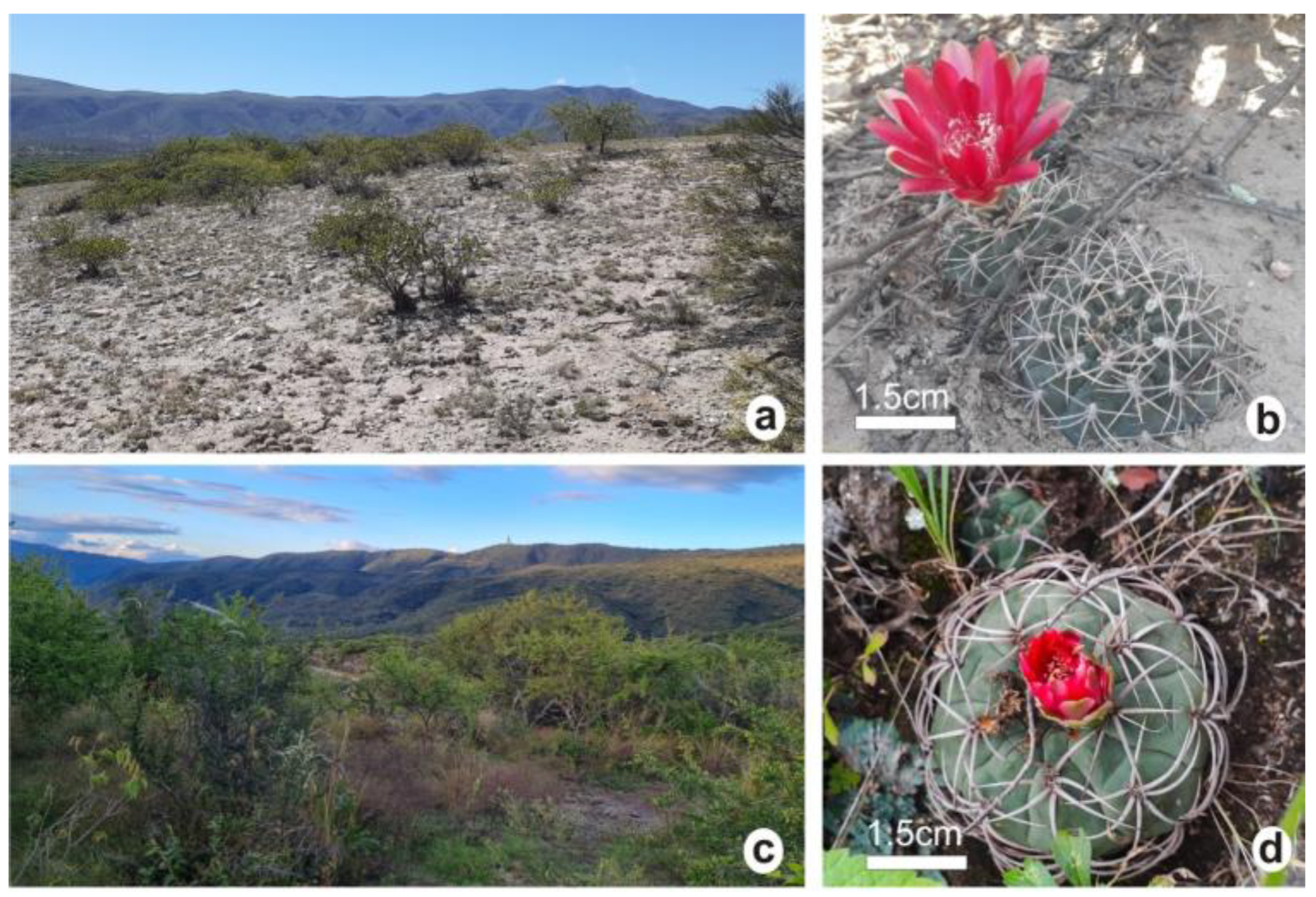
:1. Introduction
2. Results
2.1. Plant Morphology
2.2. Photosynthetic Pigments
2.3. Biomass Distribution
2.4. Ectomycorrhizae
2.5. Root Anatomy
2.6. Stem Anatomy
2.7. Spine Anatomy
2.8. Phenolic Compounds
3. Discussion
4. Materials and Methods
4.1. Habitat Traits of Species under Study
4.2. Plant Material
4.3. Morphological Characterization, Photosynthetic Pigments, and Biomass
4.4. Ectomycorrhizae Morphology
4.5. Anatomical Analysis
4.6. Transmission (TEM) and Scanning (SEM) Electron Microscopy
4.7. Soluble Phenolic Compound Determination
4.8. Statistical Analysis
5. Conclusions
Author Contributions
Funding
Data Availability Statement
Acknowledgments
Conflicts of Interest
References
- Trevisson, M.; Perea, M. Cactus del Oeste de Argentina, 1st ed.; L.O.L.A.: Buenos Aires, Argentina, 2016; pp. 6–7. [Google Scholar]
- Zuloaga, F.O.; Belgrano, M.J.; Zanotti, C.A. Actualización del Catálogo de las Plantas Vasculares del Cono Sur. Darwiniana 2019, 7, 208–278. [Google Scholar] [CrossRef]
- Demaio, P.H.; Barfuss, M.H.J.; Kiesling, R.; Till, W.; Chiapella, J.O. Molecular phylogeny of Gymnocalycium (Cactaceae): Assessment of alternative infrageneric systems, a new Subgenus, and trends in the evolution of the genus. Am. J. Bot. 2011, 98, 1841–1854. [Google Scholar] [CrossRef] [PubMed] [Green Version]
- Perotti, S.B.; Aliscioni, N.L.; Delbón, N.E.; Perea, M.; Hammann, A.; Gurvich, D.E. Biomass partitioning and morphoanatomical traits of six Gymnocalycium (Cactaceae) species occurring along a precipitation gradient. Diversity 2022, 14, 749. [Google Scholar] [CrossRef]
- IUCN. The IUCN Red List of Threatened Species. Available online: https://www.iucnredlist.org/search/list?query=gymnocalycium&searchType=species (accessed on 15 July 2023).
- Schulze, E.D.; Beck, E.; Buchmann, N.; Clemens, S.; Müller-Hohenstein, K.; Scherer-Lorenzen, M. Plant Ecology, 2nd ed.; Springer: Berlin/Heidelberg, Germany, 2019; p. 926. [Google Scholar]
- Taiz, L.; Zeiger, E.; Moller, I.M.; Murphy, A. Fundamentals of Plant Physiology, 1st ed.; Oxford University Press: New York, NY, USA, 2018; p. 561. [Google Scholar]
- Carrillo-Garcia, A.; Leon de la Luz, J.L.; Bashanl, Y.; Bethlenfalvay, G.J. Nurse plants, mycorrhizae, and plant establishment in a disturbed area of the Sonoran Desert. Restor. Ecol. 1999, 7, 321–335. [Google Scholar] [CrossRef] [Green Version]
- Fracchia, S.; Aranda, A.; Gopar, A.; Silvani, V.; Fernandez, L.; Godeas, A. Mycorrhizal status of plant species in the Chaco Serrano Woodland from central Argentina. Mycorrhiza 2009, 19, 205–214. [Google Scholar] [CrossRef]
- Loza-Cornejo, S.; Aparicio-Fernández, X.; Patakfalvi, R.J.; Rosas-Saito, G.H. Caracteres anatómicos y fitoquímicos del tallo y raíz de Mammillaria uncinata (Cactaceae). Acta Bot. Mex. 2017, 120, 21–38. [Google Scholar] [CrossRef] [Green Version]
- Garcia, J.S.; Scremin-Dias, E.; Soffiatti, P. Stem and root anatomy of two species of Echinopsis (Trichocereeae: Cactaceae). Rev. Mex. Biodivers. 2012, 83, 1036–1044. [Google Scholar] [CrossRef]
- Soffiatti, P.; Angyalossy, V. Stem anatomy of Cipocereus (Cactaceae). Bradleya 2003, 21, 39–48. [Google Scholar] [CrossRef]
- Herrera-Cárdenas, R.; Terrazas, T.; Loza-Cornejo, S. Anatomía comparada del tallo y de la raíz de las especies del género Neoevansia Marshall (Cactaceae). Bol. Soc. Bot. México 2000, 67, 5–16. [Google Scholar] [CrossRef] [Green Version]
- Gebauer, R.; Řepka, R.; Šmudla, R.; Mamoňová, M.; Ďurkovič, J. Anatomical and morphological spine variation in Gymnocalycium kieslingii subsp. castaneum (Cactaceae). PhytoKeys 2016, 69, 1–15. [Google Scholar] [CrossRef] [Green Version]
- Stone-Palmquist, M.E.; Mauseth, J.D. The Structure of enlarged storage roots in Cacti. Int. J. Plant Sci. 2002, 163, 89–98. [Google Scholar] [CrossRef]
- Brundrett, M.C. Mycorrhizal associations and other means of nutrition of vascular plants: Understanding the global diversity of host plants by resolving conflicting information and developing reliable means of diagnosis. Plant Soil 2009, 320, 37–77. [Google Scholar] [CrossRef]
- Liu, Y.; Li, X.; Kou, Y. Ectomycorrhizal Fungi: Participation in nutrient turnover and community assembly pattern in forest ecosystems. Forests 2020, 11, 453. [Google Scholar] [CrossRef] [Green Version]
- Cumming, J.R.; Zawaski, C.; Desai, S.; Collart, F.R. Phosphorus disequilibrium in the tripartite plant ectomycorrhiza-plant growth promoting rhizobacterial association. Soil Sci. Plant Nutr. 2015, 15, 464–485. [Google Scholar] [CrossRef]
- Usman, M.; Ho-Plágaro, T.; Frank, H.E.R.; Calvo-Polanco, M.; Gaillard, I.; Garcia, K.; Zimmermann, S.D. Mycorrhizal symbiosis for better adaptation of trees to abiotic stress caused by climate change in temperate and boreal forests. Front. For. Glob. Chang. 2021, 4, 742392. [Google Scholar] [CrossRef]
- Kakouridis, A.; Hagen, J.A.; Kan, M.P.; Mambelli, S.; Feldman, L.J.; Herman, D.J.; Weber, P.K.; Pett-Ridge, J.; Firestone, M.K. Routes to roots: Direct evidence of water transport by arbuscular mycorrhizal fungi to host plants. New Phytol. 2022, 236, 210–221. [Google Scholar] [CrossRef]
- Sutela, S.; Niemi, K.; Edesi, J.; Laakso, T.; Saranpää, P.; Vuosku, J.; Mäkelä, R.; Tiimonen, H.; Chiang, V.L.; Koskimäki, J.; et al. Phenolic compounds in ectomycorrhizal interaction of lignin modified silver birch. BMC Plant Biol. 2009, 9, 124. [Google Scholar] [CrossRef] [Green Version]
- Perrotta, V.G.; Arambarri, A.M. Cladodes anatomy of Opuntia (Cactaceae) from the province of Buenos Aires (Argentina). Bol. Soc. Argent. Bot. 2018, 53, 345–357. [Google Scholar] [CrossRef] [Green Version]
- Males, J.; Griffiths, H. Stomatal Biology of CAM Plants. Plant Physiol. 2017, 174, 550–560. [Google Scholar] [CrossRef] [Green Version]
- Buchanan, B.B.; Gruissem, W.; Jones, R.L. Biochemistry and Molecular Biology of Plants, 2nd ed.; Wiley-Blackwell: Hoboken, NJ, USA, 2015; p. 1280. [Google Scholar]
- Loza-Cornejo, S.; Terrazas, T. Epidermal and hypodermal characteristics in North American Cactoideae (Cactaceae). J. Plant Res. 2003, 116, 27–35. [Google Scholar] [CrossRef]
- Faigón, A.; Galati, B.; Rosenfeldt, S.; Kiesling, R. Epidermal Characters of Pterocactus (Opuntioideae, Cactaceae). Haseltonia 2010, 16, 57–66. [Google Scholar] [CrossRef]
- Bobba, M.E. Clima de Montaña. El caso del Valle del Suncho Campo del Pucará; Departamento de Geografía, Facultad de Filosofía y Letras, Universidad Nacional de Tucumán: San Miguel de Tucumán, Argentina. Available online: http://observatoriogeograficoamericalatina.org.mx/egal6/Procesosambientales/Climatologia/861.pdf (accessed on 25 June 2023).
- Patané Aráoz, C.J. Estudios arqueológicos en el Pukará del Aconquija (Dpto. Andalgalá, Prov. de Catamarca). In Aconquija. un Pueblo Originario, Historia y Belleza en un Punto Común; Alaniz, H., Ed.; Municipalidad de Aconquija: Andalgalá, Argentina, 2010; pp. 153–158. [Google Scholar]
- Palmieri, C.N.; Olmos, L.R.; Quiroga, A.; de la Orden, E.; Carma, M.I. Caracterización hidroclimática de siete localidades del Departamento Ambato. Provincia de Catamarca. Argentina. CIZAS 2005, 6, 7–17. [Google Scholar]
- Wellburn, A.R. The spectral determination of chlorophylls a and b, as well as total carotenoids, using various solvents with spectrophotometers of different resolution. J. Plant Physiol. 1994, 144, 307–313. [Google Scholar] [CrossRef]
- Agerer, R. Anatomical characteristics of identified ectomycorrhizas: An attempt towards a natural classification. In Mycorrhiza. Structure, Function, Molecular Biology and Biotechnology, 2nd ed.; Varma, A., Hock, B., Eds.; Springer: Berlin, Germany, 1999; pp. 633–682. [Google Scholar]
- D’Ambrogio de Argüeso, A. Manual de Técnicas en Histología Vegetal; Hemisferio Sur: Buenos Aires, Argentina, 1986; p. 83. [Google Scholar]
- Zarlavsky, G.E. Histología Vegetal: Técnicas Simples y Complejas; Sociedad Argentina de Botánica: Buenos Aires, Argentina, 2014; p. 198. [Google Scholar]
- Dilcher, D.L. Approaches to the identification of angiosperm leaves remains. Bot. Rev. 1974, 40, 1–157. [Google Scholar] [CrossRef]
- Karnovsky, M.J. A formaldehyde glutaraldehyde fixative of high osmolality for use in electron microscopy. J. Cell Biol. 1965, 27, 137–138. Available online: http://www.jstor.org/stable/1604673 (accessed on 15 April 2023).
- Heslop-Harrison, Y.; Heslop-Harrison, J. The digestive gland of pinguicula: Structure and cytochemistry. Ann. Bot. 1981, 47, 293–319. [Google Scholar] [CrossRef]
- Venable, J.R.; Coggeshall, R. A simplified lead citrate stain for use in electron microscopy. J. Cell Biol. 1965, 25, 407–408. [Google Scholar] [CrossRef]
- Singleton, V.; Orthofer, R.; Lamuela-Raventós, R. Analysis of total phenols and other oxidation substrates and antioxidants by means of Folin-Ciocalteu reagent. Methods Enzymol. 1999, 299, 152–178. [Google Scholar] [CrossRef]
- Woisky, R.; Salatino, A. Analysis of propolis: Some parameters and procedures for chemical quality control. J. Apic. Res. 1998, 37, 99–105. [Google Scholar] [CrossRef]


| Organs and Stomata | G. marianae | G. oenanthemum |
|---|---|---|
| Stem diameter (mm) | 28.30 ± 1.90 b | 31.48 ± 2.67 a |
| Stem length (mm) | 32.87 ± 5.77 a | 27.30 ± 4.82 b |
| Root length (mm) | 57.74 ± 12.79 a | 64.26 ± 14.89 a |
| Stoma length (µm) | 101.2 ± 5.0 a | 88.4 ± 5.3 b |
| Stoma width (µm) | 79.8 ± 3.5 a | 63.7 ± 4.4 b |
| Stomatal density (mm2) | 40.4 ± 2.7 a | 19.6 ± 1.5 b |
| Photosynthetic pigments (µg g−1 FW) | ||
| Chlorophyll a | 405.35 ± 3.44 a | 275.98 ± 5.75 b |
| Chlorophyll b | 140.26 ± 6.20 a | 107.37 ± 18.51 b |
| Chl a/Chl b | 2.89 ± 0.19 a | 2.57 ± 0.15 b |
| Carotenoids | 73.25 ± 1.86 a | 46.54 ± 1.94 b |
| Parameters | G. marianae | G. oenanthemum | ||||
|---|---|---|---|---|---|---|
| Stem | Spine | Root | Stem | Spine | Root | |
| FW (g) | 8.81 ± 1.78 a | 0.16 ± 0.06 a | 0.85 ± 0.30 a | 9.74 ± 2.02 a | 0.20 ± 0.08 a | 0.64 ± 0.21 b |
| DW (g) | 1.06 ± 0.20 a | 0.15 ± 0.05 a | 0.18 ± 0.06 a | 0.87 ± 0.15 b | 0.18 ± 0.07 a | 0.11 ± 0.03 b |
| DWD (%) | 76.26 ± 2.40 a | 10.79 ± 0.50 b | 12.95 ± 1.20 a | 75.00 ± 1.98 a | 15.52 ± 0.26 a | 9.48 ± 1.32 b |
| DW/FW | 0.12 ± 0.01 a | 0.92 ± 0.02 a | 0.21 ± 0.03 a | 0.09 ± 0.01 b | 0.88 ± 0.03 b | 0.18 ± 0.02 b |
| Water content (%) | 87.72 ± 1.91 b | 8.36 ± 3.05 b | 78.89 ± 2.70 b | 90.92 ± 1.62 a | 11.76 ± 3.11 a | 81.64 ± 2.96 a |
Disclaimer/Publisher’s Note: The statements, opinions and data contained in all publications are solely those of the individual author(s) and contributor(s) and not of MDPI and/or the editor(s). MDPI and/or the editor(s) disclaim responsibility for any injury to people or property resulting from any ideas, methods, instructions or products referred to in the content. |
© 2023 by the authors. Licensee MDPI, Basel, Switzerland. This article is an open access article distributed under the terms and conditions of the Creative Commons Attribution (CC BY) license (https://creativecommons.org/licenses/by/4.0/).
Share and Cite
Soto Acosta, M.E.; Perea, M.; Ruiz, A.I.; Hilal, M.; Albornoz, P.L.; Isla, M.I. Adaptative Strategies in Gymnocalycium Species (Cactaceae) and the Presence of Ectomycorrhizae Associated with Survival in Arid Environments. Plants 2023, 12, 2774. https://doi.org/10.3390/plants12152774
Soto Acosta ME, Perea M, Ruiz AI, Hilal M, Albornoz PL, Isla MI. Adaptative Strategies in Gymnocalycium Species (Cactaceae) and the Presence of Ectomycorrhizae Associated with Survival in Arid Environments. Plants. 2023; 12(15):2774. https://doi.org/10.3390/plants12152774
Chicago/Turabian StyleSoto Acosta, María E., Mario Perea, Ana I. Ruiz, Mirna Hilal, Patricia L. Albornoz, and María I. Isla. 2023. "Adaptative Strategies in Gymnocalycium Species (Cactaceae) and the Presence of Ectomycorrhizae Associated with Survival in Arid Environments" Plants 12, no. 15: 2774. https://doi.org/10.3390/plants12152774